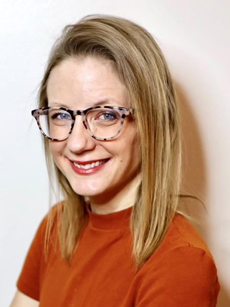
Indianapolis faculty: Rachel Surowiec
Purdue University in Indianapolis launched on July 1, 2024, ending a long-standing collaboration between Purdue University and Indiana University at what was called Indiana University-Purdue University Indianapolis (IUPUI). Purdue University in Indianapolis is an expansion of Purdue West Lafayette, with multiple locations throughout the city of Indianapolis. It bookends America’s Hard Tech Corridor, which extends from West Lafayette to downtown Indianapolis, and it positions our students, faculty and staff in close proximity to top businesses and leading industries, providing new and unique executive education and internship opportunities.
Among the Purdue faculty Indianapolis is Rachel Surowiec, assistant professor in Weldon School of Biomedical Engineering (she is also an adjunct assistant professor of radiology and imaging sciences at the nearby Indiana University School of Medicine). Surowiec is taking full advantage of the Indianapolis location to work with at least five world-class medical centers within a 10-minute walk of the campus to further her research linking multiple subject matter disciplines via engineering and computational innovation to help resolve the most vexing clinical challenges in bone fragility.
What is your research focus? What investigative avenue are you pursuing?
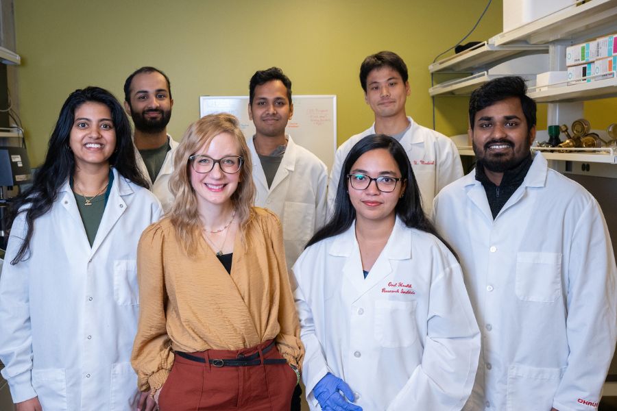
My research focuses on advancing imaging-based biomarkers to detect skeletal fragility earlier, uncover its underlying mechanisms, and enable more personalized treatment. I direct the Quantitative Biomedical Imaging and Spectroscopy Lab at Purdue University, where our interdisciplinary team bridges imaging science, deep and machine learning, musculoskeletal biology, and mechanics through engineering and computational innovation.
I love imaging —both its complexity and its potential — and am driven to maximize the information it can provide to uncover new angles on long-standing clinical challenges, from earlier fracture prediction to more targeted therapeutic guidance. I also love mentoring and team science — and I’m so grateful to work with a fantastic group of five PhD students who honestly push the science in every way to be better.
One of our core efforts centers on high-resolution peripheral quantitative computed tomography (HR-pQCT) and radiomics, an image-based “-omics” approach that extracts hundreds of quantitative features (e.g., texture, intensity, shape) from scans — similar to how genomics reveals complexity from DNA. We apply radiomics to HR-pQCT images of bone and muscle in chronic kidney disease (CKD), where fracture risk is strikingly high yet poorly predicted, and in individuals undergoing pharmacologic weight-loss therapies like Ozempic, to better understand how rapid weight loss affects musculoskeletal integrity and to identify biomarkers of resilience versus decline. This work is powered by an exceptional team of Weldon School PhD students and undergraduate researchers from across Purdue Engineering, whose energy and expertise has absolutely accelerated this work.
We are also investigating bone water as a key contributor to skeletal fragility. Using Fourier-transform near-infrared spectroscopy and thermogravimetric analysis, we quantify changes in matrix water compartments. These findings are being translated to the imaging scale using ultrashort echo time and rosette petal trajectory MRI to spatially map bone water in vivo. In parallel, we are developing motion correction methods for HR-pQCT and advanced MRI to reduce artifacts, preserve image quality, and increase clinical and research usability.
Ultimately, our goal is to move beyond density alone and build biology-informed imaging diagnostics that provide earlier, more accurate and actionable insights for musculoskeletal health.
How vital is it for you to be in Indianapolis?
Being in Indianapolis is everything. When I walk out the door of my lab, I’m within a 10-minute walk of at least five world-class medical centers. That level of proximity has made collaboration seamless and deeply rewarding. I can build strong, lasting relationships with clinical colleagues through in-person conversations, regular meetings and shared access to patient populations.
For example, together with Dr. Chris Newman (the musculoskeletal pediatric radiologist at Riley Hospital for Children and adjunct in BME), Dr. Erik Imel (IU School of Medicine endocrinologist) and Dr. Stuart Warden (IU School of Health and Human Sciences and director of the Function, Imaging, and Testing Core team at IU School of Medicine), we’ve launched an Indiana Center for Musculoskeletal Health-funded pilot study on X-linked hypophosphatemia, a rare and severe skeletal disease. We’re prospectively enrolling patients and acquiring a new experimental MRI sequence to identify bone imaging biomarkers that could detect X-linked hypophosphatemia-related fragility — something that current imaging tools like bone mineral density simply can’t do well.
It’s a highly multidisciplinary, multi-institutional team, yet we’re all within walking distance. That kind of physical and intellectual proximity allows us to meet often, iterate quickly and move the science forward with access to a rare and highly motivated patient population. Physicians like Dr. Imel are deeply committed to improving outcomes for their patients, and with engineering embedded here in Indy, we’re able to bring the technical solutions that make a real difference. It’s been a dream collaboration.
For my students, this environment is just as powerful. They have direct access to clinical imaging workflows and get to witness real-world problems that need engineering solutions. One great example is Farhan Sadik, one of my PhD students, who is working closely with the FIT Core. One major challenge with high-resolution HR-pQCT imaging is motion — any movement during the scan significantly blurs bone microarchitecture, leading to compromised data in up to 20% of cases. This results in costly rescans, unnecessary radiation exposure (albeit minimal), and workflow inefficiencies. Farhan, along with a team of undergraduate researchers he mentors, is developing a solution that simulates motion artifacts and corrects them using AI. This work has already progressed toward a provisional patent filing.
As a PI, I couldn’t be prouder. It’s a perfect example of biomedical engineering in action — and it’s only possible because of the access and environment we have here in Indianapolis.
What is the environment like at PIN?
What I love most about this campus is the students. Their energy, curiosity and passion for discovery are absolutely infectious. They’re not only excited about research — they’re shaping it. Whether it’s engaging with local tech startups, working on cutting-edge biomedical innovations, or connecting with major industry players like Eli Lilly, the opportunities here are real and right at their fingertips. Clinical access is unparalleled — partnerships with the hospital systems and medical school are not just possible, they’re woven into the everyday research experience.
Getting students connected — tethered — to research is one of my greatest passions. And being at Purdue and located in Indianapolis makes it possible in really dynamic ways due to being embedded in an urban setting and adjacent to the medical centers.
What is your philosophy of teaching?
I love this question because my teaching philosophy is always evolving. Teaching is an incredible feedback loop — every time I step into the classroom, I learn something too. At its core, I want students to see the beauty in the fundamentals — especially in math and physics. For example, I teach a sophomore-level course on electrical circuit theory. At first glance, it might seem abstract or dry, but when you realize that these principles power devices that keep hearts beating or allow us to measure complex processes in the body — it becomes deeply human and meaningful. It’s my responsibility to bring that to life for them.
Early on, I was nervous about being asked something beyond my expertise. Now, I welcome it. When a student asks a question I haven’t considered, it’s often a sign they’ve reached that “aha” moment — when they’re genuinely wondering how or why something works. That kind of engagement is everything. I never want them to stop wondering.
At the graduate level, I’ve had the privilege of developing two courses so far: Biomedical Ultrasound and Applied Medical Image Processing and Analysis. Both are anchored in real-world data and give students hands-on experience with challenges they might encounter in research or industry. Watching them light up when they connect theory to application has been one of the most rewarding parts of my role as an educator.
What was your path like prior to present day?
Thank you for asking! I’m a first-generation college student and was the first in my family to graduate high school on time. My parents have always been my biggest cheerleaders. When they saw how much I loved school, they made a life-changing decision — they went back to night school and earned their own high school diplomas while I was in my early teens. They wanted to lead by example, and their courage opened up a world of possibilities for me.
I went to Ball State University, where I also played tennis and fell in love with biomechanics. That was my first big “aha” moment — I found the biomechanics lab, caught the research bug and stayed for a thesis-based master’s. It was a formative experience. I got to work on FEA models to predict stress fractures and ran motion capture studies with pediatric patients with cerebral palsy, pre- and post-intervention. I watched how physicians used the data I helped collect to guide real clinical decisions. That was powerful.
I next joined the Imaging Research Department at the Steadman Philippon Research Institute in Vail, Colorado. We worked closely with both academia and industry, especially Siemens, using quantitative MRI to identify biomarkers for post-traumatic osteoarthritis. One of our sequences even became part of the standard imaging protocol for all patients at the clinic — a true bench-to-bedside win. We also began exploring automated segmentation and our method and subregions for analysis was incorporated into a software package; all incredibly informative experiences working alongside industry in a research setting.
Wanting to grow in mentorship, team building and grantsmanship, I applied to the NSF Graduate Research Fellowship Program, received it and pursued my PhD in biomedical engineering at the University of Michigan with a concentration on biomedical imaging, focusing on quantitative MRI and biomarker development in bone. I trained under Dr. Ken Kozloff in orthopaedic surgery and had the space to swing big with ideas. I then joined the Center for Molecular Imaging in the Department of Radiology at Michigan for a postdoc — this time pivoting to molecular imaging and brain cancer, studying stem-like cells in diffuse intrinsic pontine glioma, a universally fatal pediatric brain tumor.
Training outside the musculoskeletal space deeply shaped how I think about imaging today. I later completed a second postdoc at IU School of Medicine in both the Department of Anatomy, Cell Biology & Physiology and the Weldon School of Biomedical Engineering. That’s when I returned to bone, imaging and chronic kidney disease — and I fell in love with the campus, the people and the research environment. I knew I had found my research home. When a position opened in BME, I gave it everything I had. It was the best decision I’ve made.
I also want to share something I find important, especially for trainees. I returned for my PhD after nearly five years in the field, and for me, that was absolutely the right decision. I also started my family during my training — my first son turned one right around my defense, I had my second son during my postdoc and my daughter during my faculty role. Seeing researchers raise families while leading outstanding programs has meant everything to me. I’m passionate about improving academic culture and policy to support caregivers, especially during the early parenting years and later stages of life when many of us care for aging parents. It's a big part of who I am and how I approach my career.
What about your future? What are you looking to accomplish in this field?
I want to fundamentally change how we detect and understand bone fragility — shifting the focus from solely identifying risk to also identifying resilience. Why do some individuals maintain skeletal health despite disease or physiological stress, while others do not? This question drives much of my work. Whether it’s rapid weight loss or chronic kidney disease, we need better tools to identify who is most vulnerable and why.
My lab is developing imaging-based biomarkers that go beyond bone mineral density to include components like collagen and water — water alone accounts for nearly 20% of cortical bone by volume and plays a key role in bone mechanics. We've found that matrix-bound water is modulated during disease progression and may represent a novel therapeutic target. Our work leverages advanced imaging techniques that either avoid radiation or maximize information from existing modalities.
I also care deeply about staying connected to my scientific community. I’m active in the American Society for Bone and Mineral Research, where I currently serve on the Women in Bone Committee. It’s been a vital part of my career development, and I’m committed to giving back to the field that helped shape me.
Another exciting area is the application of machine and deep learning in bone imaging. This is just beginning to take hold in our field, and I’m excited that our lab is helping push aspects of this field forward. We’ve been applying radiomics, which has already revolutionized cancer imaging, and we believe it holds similar promise for musculoskeletal health. Our goal is to uncover patterns and features invisible to the human eye but deeply informative about tissue composition and structural integrity.
Lastly, I want to mentor the next generation of researchers. If years from now my former students are leading their own labs, innovating in industry, or founding impactful startups — that will be one of my greatest accomplishments. Their success means more to me than any personal accolade, and I feel lucky every day to play a part in their journey.
Might you share with us a little window into your personality; some distinctive trait, habit of mind, hobby/pursuit outside work…?
Outside the walls of my lab, my favorite role is being a mom. I have two boys (ages 7 and 4) and a 1-year-old daughter who keep me on my toes. Right now, we’re deep in our Pokémon era — collecting cards, watching the original series and playing Pokémon Go together. On weekends, you might find us hunting down the latest deck release or walking around the Indy campus, which is full of PokéStops (my kids now expect me to “stock up on Poké Balls” before I head home from work!).
My oldest, Sebi, and I have recently started learning to play the drums together — he’s already better than I am, but I’m keeping up as best I can. I also love running (slowly!) and have developed a full-blown obsession with gardening. Obsession doesn’t necessarily mean skill, but I’m giving it my best.