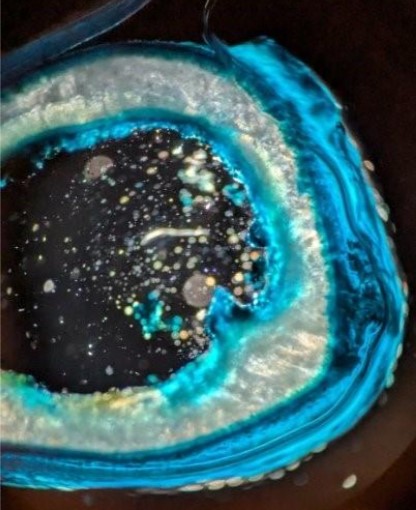
Art in Research

Science is an art, but not all scientists are artists.

That said, we purposefully encourage our Biomedical Engineering students to express their creativity while showcasing their research.

Each fall, students submit images—photographs, micrographs, illustrations, visualizations and renderings—depicting their science, along with a description of how the image was created and how it represents the importance of research in their field. Art in Research is intended to be a record of striking visual qualities and a lens into the science, to be shared with fellow researchers and the general public alike.

A Moment in Time

Submitted by: Luke Shinneman

Category: Microscopy

Contributions: Leo Green

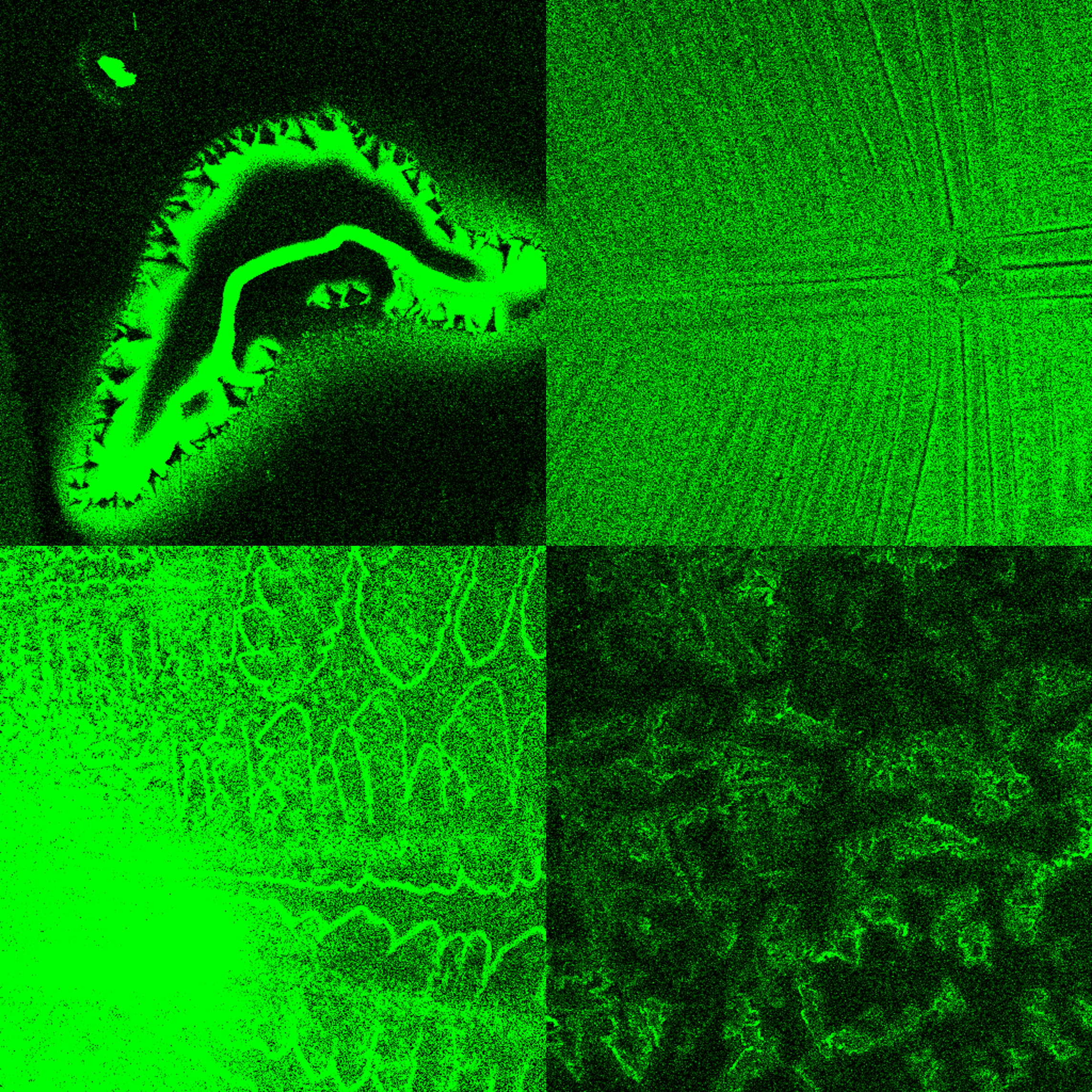
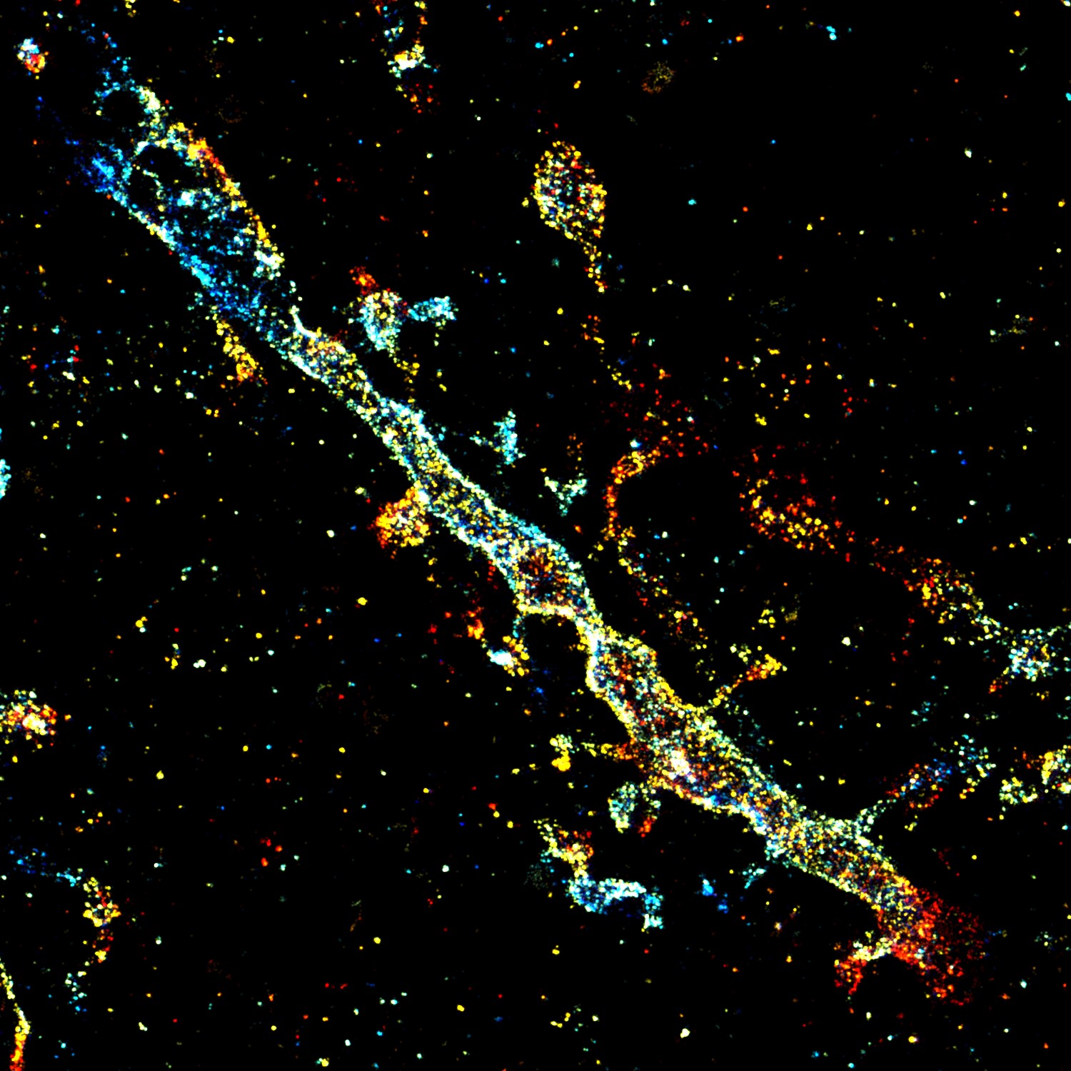
Creation: Microbes from the soil in West Lafayette were cultured on the surface of gelatin-halide film prints.

Caption: This photograph was taken on my 1975 Yashica film camera on a trip to the Galápagos Islands of Ecuador. Prior to inkjet printing in the 1990’s, photographs were printed on gelatin-halide emulsions like these. Taking advantage of microbes’ ability to break down and recycle large proteins, we cultured bacteria and fungi from the soil of West Lafayette on the surface of these prints, and captured their final progress in a moment in time..


A Purdue logo

Submitted by: Van Thuy Duong

Category: Microscopy

Creation: We attempted to achieve spatially dynamic stiffening and softening within the same dSIS-NB-only (our novel bimaterial) gel using micropatterning with the LumenX bioprinter. A simple Purdue University logo was designed using the TinkerCAD website. The logo consisted of two parts: a larger ‘P’ designated for stiffening and a smaller ‘P’ for softening, which overlaid the larger ‘P’. The pre-formed soft gel was stiffened for 60 seconds with 0.5 mM tartrazine, and the larger ‘P’ appeared on the gel in a darker color. The gel was then washed and incubated with 5 mM Rh-PEGSH (red fluorescent molecules) and 6 mM LAP (photo initiator) before exposure with the smaller ‘P’ for 60seconds with 0.5 mM tartrazine. The smaller ‘P’ overlaid the larger ‘P’ to form a Purdue University logo. The red area was initially soft, then stiffened, and then softened again. The hydrogel was imaged using a 3D fluorescent confocal microscope (BC43) using a 2X objective.

Bloom of the Human Gut Assembloids-on-a-Chip

Submitted by: Jong-Keun Sim

Category: Microscopy

Contributors: Leandra Suter and Estelle Park

Creation: The human assembloids-on-a-chip platform combines stem cell biology with microengineering to generate complex intestinal tissue structures in vitro. By guiding the fusion of multiple human intestinal organoids and exposing them to controlled soluble gradients, the system promotes the emergence of budding morphologies that closely mimic the crypt–villus architecture of the intestine. This innovation allows researchers to model spatial organization and signaling dynamics of the tissue with unprecedented precision, advancing studies in development, disease modeling, and drug discovery.

Caption: A confocal micrograph of a human gut assembloid formed within a microengineered chip. At the base, fluorescently labeled microchannels in red and green highlight the device structure. Above, the fused intestinal organoids form a large budding tissue that mimics the crypt–villus structure, stained with phalloidin (magenta, actin cytoskeleton) and DAPI (green, nuclei). The image resembles a blooming flower, visually capturing how engineered platforms can recreate the budding architecture of the intestine in vitro. Gut assemblage size: 1747 μm. Device dimensions: 25 mm (width) × 32 mm (length) × 9.6 mm (height).

Fracture in a Mouse Bone

Submitted by: Olivia White

Category: Microscopy

Contributors: Rachel Kohler, Katelynn Gallagher, Dyann Segvich, Karthik Rajesh and Joey Wallace

Creation: Scanning Electron Microscopy

Caption: This piece shows the cross-section of a femur from a mouse after fracture toughness testing. In fracture toughness testing, the bone is notched and broken, then imaged using a scanning electron microscope. In this image, the notch is the smooth surface at the bottom of the bone. Stable growth of the crack during testing is represented by the striations in the bone surface, extending from the notch. Measuring the angle from the center of the bone cross-section to the notch edge and to the end of stable crack growth are each measured. These values can then be used to calculate properties of fracture toughness. Fracture toughness measures can help researchers understand how different treatments, diseases, etc., may impact the fracture resistance of a bone.

Larvae dancing flow

Submitted by: Shulan Xiao

Category: Anatomy & Physiology

Contributors: Sanket Samal, Tyler Pikes, Qing Deng, Jianguo Mei and Krishna Jayant

Creation: The swimming trajectories of zebrafish larvae during c-turning behavior, captured by a high-speed camera at 750Hz, labeled with Deeplabcut and visualized with Matlab.

Caption: The swimming trajectories of zebrafish larvae under external sensory stimuli - with injectable tissue-like electrodes grown in their bodies! Fun fact: the ultrasoft grown optoelectrical interface doesn't affect their motion, and they can turn around within ~20ms, as fast as other larvae.

Mother of the Cosmos

Submitted by: Tuba Marjan

Category: Microscopy

Creation: Fibroblast cells were cultured on hybrid 2D hydrogels and stained to visualize actin filaments and nuclei, then imaged using fluorescence microscopy. The images were artistically processed to enhance contrast and highlight the structural details of the cells.

Caption: The actin cytoskeleton of fibroblast cells forms intricate networks around brightly stained nuclei. This microscopic view subtly mirrors the patterns of constellations observed in the night sky.

Neurons in the Vascular Jungle

Submitted by: Doohyeong Jang

Category: Microscopy

Creation: 2P microscopy with some post-processing

Caption: This piece captures the delicate yet complex relationship between neurons and the dense network of blood vessels that sustains them. The glowing green dots represent neuronal bodies, scattered like fireflies in a night forest. The red filaments, twisting and branching across the canvas, evoke a jungle canopy of vessels, emphasizing the intricate vascular web that both nourishes and conceals the neurons. The deep blue background creates a dreamlike environment, situating the viewer inside a hidden microcosm of the brain. By presenting the microscopic world in vibrant colors and organic forms, the image blurs the boundary between science and art, inviting us to see the brain not only as a site of cognition but also as a living landscape of connection, support, and mystery. This is AI AI-created description and I agree with this :D...

Scientific Muses

Submitted by: Nissa Larson

Category: Hand Drawn

Creation: Digital art.

Caption: Model organisms are the foundation of scientific discovery in the biological sciences. Three key species are shown - Drosophila melanogaster (fruitfly), Danio rerio (zebrafish), and Mus musculus (mouse).

Sprouting Through the Matrix

Submitted by: Nicole Mertens

Category: Microscopy

Contributors: Jun KIm

Creation: Spheroids from hMSCs and HUVECs were cultured inside a cell-mediated degradable hydrogel.

Caption: This piece captures the dynamic process of spheroid sprouting within a degradable bulk hydrogel, highlighting how cells interact and remodel their extracellular matrix.


The Architecture of Thought

Submitted by: Cheng Bi

Category: Microscopy

Creation: Super-resolution reconstruction of a mouse brain sample injected with Brainbow virus, immunolabeled with anti-GFP antibody conjugated to AF647, and imaged using a biplane system.

Caption: This artwork highlights the dendritic spine structures of a mouse neuron, which often appear blurred under conventional imaging. Through Brainbow labeling, anti-GFP-AF647 staining, and biplane super-resolution microscopy, these fine synaptic details are resolved with exceptional clarity.

The Forward Model

Submitted by: Hammad F. Khan

Category: Rendering

Creation: Adobe Illustrator, ImageJ, two-photon microscopy

Caption: Our limbs are endowed with neural motifs that enable exquisite control of movements. In the absence of idealized movements, we seek to restore them. This piece represents the intersection of biotechnology and neuroscience in an effort to shape neural dynamics supporting our intent to move through the world.


The Frozen Flow

Submitted by: Cortland Johns

Category: Microscopy

Creation: Scanning Electron Microscopy

Caption: Polyhedrocytes (compressed/malformed red blood cells) found in intraluminal thrombus formation in a mouse with an abdominal aortic aneurysm. Scale bar: 20 um

Vascular network within natural hydrogel

Submitted by: Van Thuy Duong

Category: Microscopy

Contributors: Ngoc Ha Luong

Creation: Human endothelial cells were encapsulated within a 3D photo-responsible decellularized extracellular matrix hydrogel and cultured for 4 days. The cells migrated and formed a functional interconnected vascular network within the hydrogel. The network was immunostained with Ve-Cadherin (green), F-Actin (red) and DAPI (blue) signal then imaged with BC43 3D fluorescent confocal microscope with a 20X objective.

Caption: Human endothelial cells proliferated and formed a functional vascular network within a 3D photo-responsible decellularized extracellular matrix hydrogel after 4 days of culture.

Waves in Bone

Submitted by: Ishita Mukadam

Category: Microscopy

Creation: Cortical bone slices were fixed, sectioned, and stained with Alcian blue to visualize glycosaminoglycan (GAG) content. Imaging was performed using a Zeiss brightfield microscope.

Caption: This image shows a rat tibia bone slice stained with Alcian blue under a brightfield microscope. The stain highlights glycosaminoglycans(GAGs) within the bone matrix, which play a crucial role in regulating bone hydration and structural integrity. By staining the bone slices, we can visually quantify the bone matrix, which is valuable in the context of biomarker chronic kidney disease (CKD).

World's smallest 3D printed P

Submitted by: Om Kolhe

Category: Device Design

Creation: This is a nanoscale 3D printed Purdue P. This was created using the Exaddon CERES 3D printer. The P is just 100um tall. The image was taken using a Hitachi SEM.

Yin and Yang

Submitted by: Ngoc Ha Luong

Category: Microscopy

Creation: Confocal microscopy of 3D biofabricated hydrogels.

Caption: A 3D printed sacrificial hydrogel (top image, the Yin) for inverse-molding an extracellular matrix hydrogel with crypts/villi topography (bottom image, the Yang).

2024

A map of neural constellations

Submitted by: Lizzy Frazier

Category: Rendering

Contributors: Megan Lipton

Creation: Two-photon (2P) calcium images of neurons were obtained from the primary visual cortex of awake mice. Images were motion-corrected usingsuite2p. The mean image of neural activity was used as the basis for mapping "neural constellations."

Caption: Just as we map the constellations of the universe, we can map the constellations of the mind. Each neuron is a star, each connection a pathway through the universe of thought.

Cellular Symphony on Hydrogel

Submitted by: Tuba Marjan

Category: Microscopy

Creation: I created this image using fluorescent microscopy to capture mesenchymal stem cells growing on a 2D hydrogel scaffold. The cells were stained with phalloidin that binds to actin filaments which form the internal structure and give shape to the cell causing them to glow bright red. The nuclei were stained with Hoechst making them glow blue. The hydrogel allowed cells to adhere and spread out. This process blends scientific observation with a visual appreciation of cellular architecture where function meets form in a dynamic expression of life.

Endoplasmic reticulum (ER) dynamics at nanoscale

Submitted by: Yue Zheng

Category: Microscopy

Contributors: Li Fang, Maryam Mahmoodi, Hao-Cheng Gao, Fan Xu, Han Zhao & Fang Huang

Creation: Photoactivation localization microscopy (PALM) imaging of a living COS-7 cell stably expressing a HaloTag on reticulum 4(Rtn4) protein stained with PA-JF549-Halo. The raw single molecule blinking data was taken on a custom-built TIRF imaging system using a 405 nm laser for photoactivation and a 561 nm laser for excitation. Data was analyzed and reconstructed using the INSPR algorithm, and the localizations in 80 seconds were plotted through a jet (64) color map.

Caption: The structural changes and fast movements of ER tubules are clearly resolved at the nanometer scale of spatial resolution and seconds level of temporal resolution. The color from blue to red indicates the actual time from 0 to 80 seconds. The scale bar at the lower right is 500 nm.

Highway to the brain

Submitted by: Sergio Martin

Category: Anatomy & Physiology

Contributors: Thejas Vishnu Ramesh

Creation: This render was created from 4D Flow MRI data acquired at the Purdue 3T GE Discovery MR750 scanner. To obtain blood flow of the carotid bifurcation, a custom-made wearable coil was used. 4D flow MRI enables three-directional measurement of blood flow velocities over the cardiac cycle. Streamlines were generated from the MRI velocity field with the software Paraview, and overlaid over the structural image, acquired with time-of-flight MRI.

Caption: The image shows the flow of blood at the right carotid artery bifurcation. This section of the carotid splits from the common carotid artery into the internal and external carotid arteries, supplying blood to the brain, face, and head. 4D flow MRI can measure blood circulation in vivo, and it can be especially useful to assess vascular health and diseases. Custom-made coils for the neck result in a higher signal, improving the acquisitions of flow and resulting in more accurate velocity fields.

Microtubule tracks in neuronal growth cone

Submitted by: Yuan Ren

Category: Microscopy

Contributors: Donghan Ma, Kristi McElmurry, Fang Huang & Daniel M. Suter

Creation: Microtubules in Aplysia growth cone imaged with tubulin immunocytochemistry. DNA-PAINT, and super-resolution microscopy using custom-made single-molecule switching nanoscopy setup.

Caption: This image shows a multitude of parallel microtubules inside a growth cone of a cultured Aplysia neuron. Microtubules are essential for organelle transport, cell division, and cell motility.

Nuclear pore complexes under super-resolution microscope

Submitted by: Hao-Cheng Gao

Category: Microscopy

Contributors: Fan Xu

Creation: Immunolabeled cells was imaged with 4Pi single-molecule localization microscopy (4Pi-SMLM) and reconstruct its ultrastructure with nanometer resolution by using 4Pi-BRAINSPOT platform.

Caption: Nuclear pore complexes are massive protein assemblies embedded in the nuclear envelope that regulate the bidirectional transport of molecules between the nucleus and cytoplasm, playing a crucial role in cellular function and gene expression. Single-molecule localization microscopy (SMLM) surpasses the diffraction limit by precisely determining the positions of individual fluorophores, while 4Pi microscopy enhances achievable resolution through interferometric optical design. Together, 4Pi-SMLM facilitates 3D imaging of proteins with nanometer precision. The color code represents the depth (Z position) of the Nup96 proteins, as indicated by the color bar.

Progression of a Murine Thoracic Aortic Aneurysm

Submitted by: Ethan Kelley

Category: Anatomy & Physiology

Contributors: Cortland Johns

Creation: EKV (EKG-gated Kilohertz Visualization) Ultrasound Imaging

Caption: EKV ultrasound imaging visualizes the progression of a murine thoracic aortic aneurysm through five-time points after the introduction of BAPN (beta-aminopropionitrile) to drinking water. BAPN inhibits collagen replacement around major arteries, causing aneurysms when administered to young mice. Major tick marks indicate millimeters. Images were collected and processed with VisualSonics Vevo3100 and Vevo Lab.

Putting the P in Purdue: Precision Magnetic Bioassembly Using Microrobot Grippers

Submitted by: Madison Howard

Category: Anatomy & Physiology

Contributors: Aaron Davis

Creation: Fluorescent cancer cells and fibroblast spheroids were spatially patterned into a "P" shape using a microrobot gripper and imaged with a fluorescent microscope.

Caption: Cell spheroids were picked and placed in a "P" shape by a wireless, magnetic microrobot gripper.

Starry Night, Cell-estial Edition

Submitted by: Claudia Benito Alston

Category: Microscopy

Contributors: Michael Deines, Madison Howard & Evelyn Nonamaker

Creation: I cultured bone Messenchymal Stem Cells (bMSCs) for 14 days, after which I fixed and stained them with an Alkaline Phosphatase labeling kit.

Caption: Staining cells with alkaline phosphatase (ALP) is commonly used to assess cell differentiation, particularly in studies involving osteogenic (bone-forming) cells. ALP is an early marker of osteoblast differentiation, meaning it is expressed when precursor cells begin to differentiate into bone-forming osteoblasts. By staining cells for ALP, we can visually confirm and quantify the activity of these cells as they progress along the osteogenic pathway.

The Human Operating System

Submitted by: Hammad Khan

Category: Rendering

Creation: Illustrator, Imagej, Device Fabrication, Neural Recordings

Caption: Created from real neural data and biophysical reconstructions of neural connections, this piece represents the next frontier for neurotechnology and physiology. Technological breakthroughs will augment dynamic models between muscles, nerves, and neurons to understand the basis for intelligence.

The Spider-Verse Within You

Submitted by: Makayla Phillips

Category: Microscopy

Contributors: Josh Sexton, Sherry Harbin & Rachel Morrison

Creation: The image was captured using a confocal microscope to analyze an immunofluorescent stained transverse muscle cross-section

Caption: An immunofluorescent laminin stain under a confocal microscope