Fracture in a Mouse Bone
Submitted by: Olivia White
Category: Microscopy
Contributors: Rachel Kohler, Katelynn Gallagher, Dyann Segvich, Karthik Rajesh and Joey Wallace
Creation: Scanning Electron Microscopy
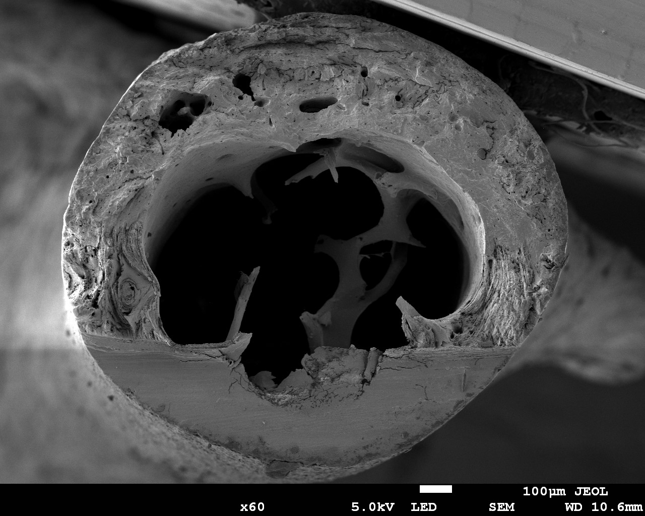
Caption: This piece shows the cross-section of a femur from a mouse after fracture toughness testing. In fracture toughness testing, the bone is notched and broken, then imaged using a scanning electron microscope. In this image, the notch is the smooth surface at the bottom of the bone. Stable growth of the crack during testing is represented by the striations in the bone surface, extending from the notch. Measuring the angle from the center of the bone cross-section to the notch edge and to the end of stable crack growth are each measured. These values can then be used to calculate properties of fracture toughness. Fracture toughness measures can help researchers understand how different treatments, diseases, etc., may impact the fracture resistance of a bone.