Fang Huang received a patent on architecture and information-based neural network

The neural networking technique utilizes Fisher information of the training dataset commonly ignored in conventional AI approaches, to pinpoint single molecule locations within cell and tissue specimens. Currently, conventional inference-based approaches to localize single molecule centers focus on dominant and recognizable features or modeling feature changes through a simplified mathematical form. Because of the difficulty of perceiving and summarizing a comprehensive PSF model, the retrieval of multiplexed information beyond 3D position from complex or high-dimensional single-molecule data remains challenging.
The deep neural network developed by Huang, named smNet, enabled multiplexed single-molecule analysis to retrieve such molecular information with high accuracy. smNet can extract three-dimensional molecule location, orientation, and wavefront distortion with precision approaching the theoretical limit, and therefore will allow multiplexed measurements through the emission pattern of a single molecule.
Key features of a single molecule such as location, orientation and wave-front distortion can now be obtained with ease. This enhanced decoupling technique allows researchers to process biological images faster than ever before.
Advantages:
- Measures at single molecule level
- Information-aware neural network training
- Real-time analysis
Potential Applications:
- Biomedical research
- Biological image processing
- Artificial Intelligence
Huang’s research has been published in Nature Methods.
Huang is the Reilly Associate Professor of Biomedical Engineering. His research interests include biomedical imaging and optics, super-resolution microscopy, cellular dynamics and structures and cell division. For additional information, visit Huang's research website: http://www.fanghuanglab.com/.
Congratulations to all Purdue University researchers across all campuses and academic disciplines, who received a patent on their intellectual property from the U.S. Patent and Trademark Office in June
Most of these innovations are available to license and bring to market. Visit the Purdue Innovates Office of Technology Commercialization’s website to learn more about these and other available innovations.
Source: Purdue Research Foundation
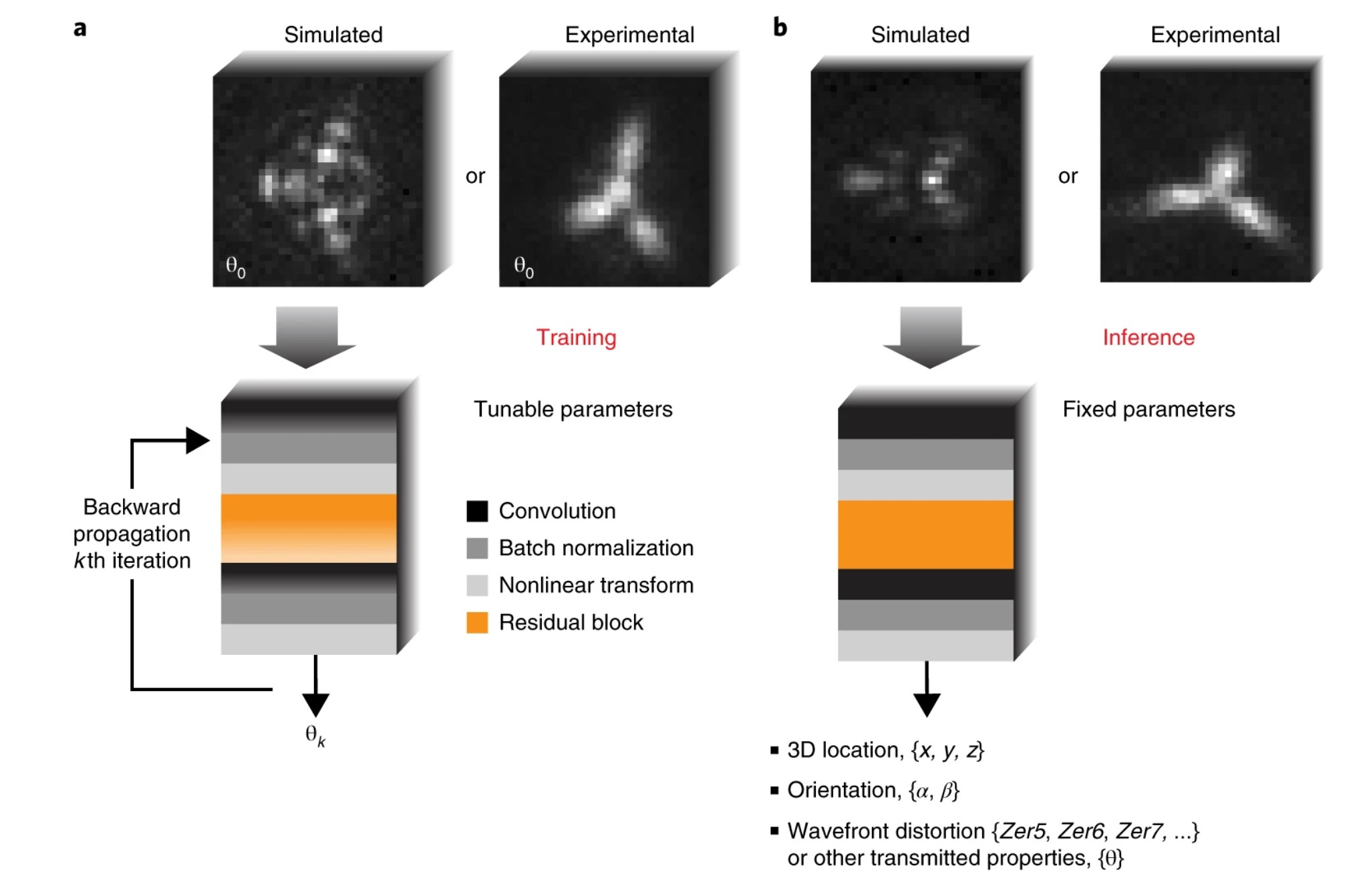
Fig 1: a, smNet training process. The example input training dataset consists of PSF images and the underlying true values of the measurements θ0. smNet takes in the training images and outputs estimations θk for the kth iteration. Iteratively, using back-propagation, the parameters in the network are updated through minimization of the designed information-limit weighted cost function. b, The estimation process with smNet. Once trained, smNet takes in a stack of PSF images and outputs the desired estimations. The simplified illustration shows four major building blocks of smNet: convolutional layer, batch normalization, nonlinear transformation, and residual block.
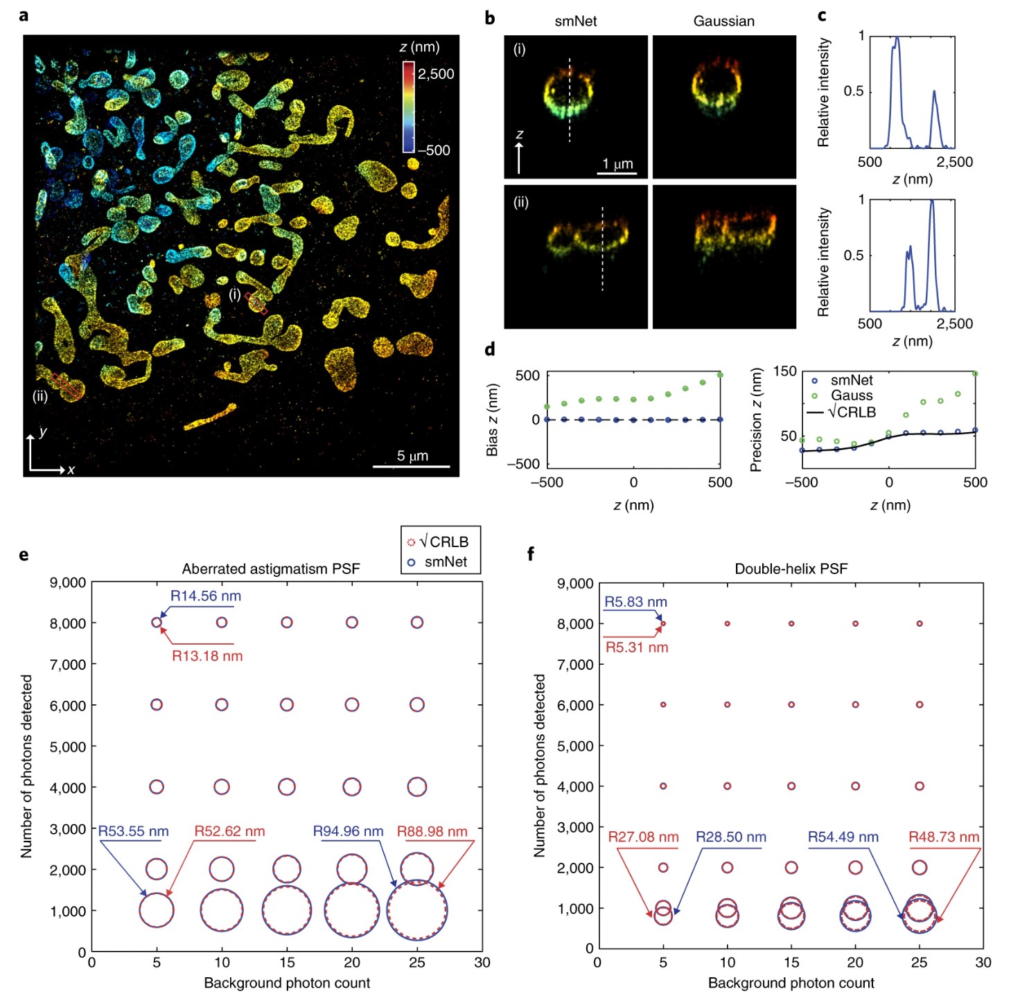
Fig 2: a, 3D SMSN reconstruction using smNet. The sample is immunolabeled TOM20 in a COS-7 cell at a depth of 12 µm from the coverslip surface. Color coding represents the relative axial position of the localized molecules. The image is representative of 9 independently repeated experiments. b, Comparison of cross-sections of regions i and ii (red boxes) in a from smNet reconstruction and Gaussian-based reconstruction (Supplementary Note 12). The data are representative of 6 independently repeated experiments. c, Intensity profiles along the dashed lines in b. The data are representative of 6 independently repeated experiments. d, Comparison of precision and accuracy for axial localization with smNet and the Gaussian-based method. Data were generated from an experimentally retrieved pupil function including aberrations at 12 µm above the coverslip. e,f, Error radii achieved at various total photon and background levels for aberrated astigmatism PSFs (e; same as in d) and double-helix PSFs (f).
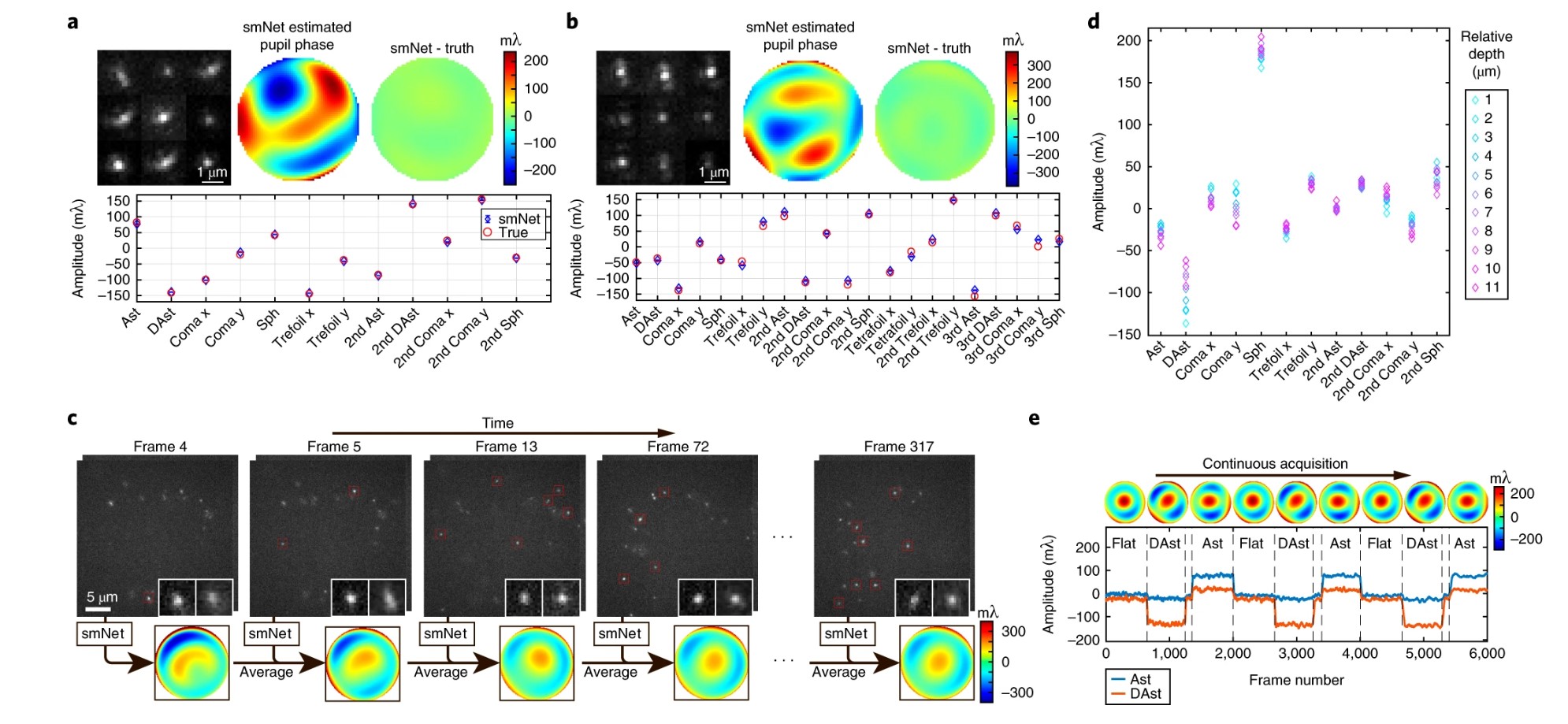
Fig 3: a,b, smNet measurement of wavefront shape composed of 12 (a) or 21 (b) Zernike modes through simulated PSFs. The amplitude of each mode was randomly sampled from –159.15 to 159.15 mλ. The nine example PSFs (upper left in each panel) were randomly picked from the first planes of the 1,000-biplane PSFs. Data are shown as mean and s.e.m. (n = 1,000). c, Illustration of smNet’s wavefront measurement based on single-molecule blinking data acquired during SMSN imaging. Subregions are enlarged to show the PSF shape in the biplane setup. d, Measurement of wavefront distortion along 11 optical sections during SMSN imaging of a whole cell. e, Tracking of dynamic wavefront changes introduced by a deformable mirror with smNet. Each dashed line represents a change in the input voltage to the deformable mirror shape, which resembles normalized Zernike polynomials. Eight experiments were repeated independently, with similar results, and additional tracking results are included in Supplementary Fig. 15 and Supplementary Data. Ast, astigmatism; DAst, diagonal astigmatism; Sph, spherical aberration.