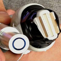
Craig Goergen receives AHA grant
An aneurysm occurs when part of an arterial wall weakens, allowing it to expand or balloon abnormally. Aneurysms can occur anywhere, but AAAs occur in the largest artery from the heart that supplies blood to rest of the body.
AAA is more common in men and in individuals aged 65 years and older. Unfortunately, AAAs are often asymptomatic until they rupture, a condition which is life-threatening. It is estimated that at least 90% of patients with a ruptured AAA die before arriving at a hospital.
In his four-year study entitled “Photoacoustic Imaging of Abdominal Aortic Aneurysms,” Goergen will study the relationship between aortic wall content, expansion rates, and rupture risk. His group will also develop an imaging system which may ultimately be used by physicians to diagnosis and treat patients with AAA.
“By studying the relationships between aortic wall composition, causes of expansion, and rupture rates associated with those variables, we may be better able to determine at an earlier stage than is currently possible which patients are at greater risk of aortic rupture.”
Collaborators on the project include: Ji-Xin Cheng, professor of biomedical engineering and professor of chemistry; Kimberly Buhman, associate professor of nutrition science, both at Purdue; and Michael Murphy, associate professor of surgery at IU School of Medicine.
|
Project Abstract: Abdominal aortic aneurysm (AAA) is a complex disease defined as a pathologic dilation of the vessel wall. The relationships between AAA wall content, expansion rate, and rupture risk have not been fully explored. Recently, whole body molecular imaging has become a research area focused on non-invasively quantifying molecular processes in vivo. One recently developed technique, vibrational photoacoustic imaging, has emerged as a new strategy for label-free molecular imaging. The purpose of this study is to develop a non-invasive photoacoustic molecular imaging system capable of characterizing and tracking vascular disease in vivo. We will first apply this technique to small animal AAA models, with a long-term goal of using these initial efforts to develop a clinical system. Our central hypothesis is that the underlying makeup of the aortic wall plays a key role in aneurysm expansion. We formed this hypothesis based on our experience imaging diseased large elastic arteries in laboratory animals and humans. In the aorta, the largest artery in the body, there is a heterogeneous mixture of lipid, collagen, elastin, and smooth muscle cells. While large aneurysms are more prone to rupture (usually defined as ≥ 5 cm), the relationships between AAA wall content, expansion rate, and rupture risk have not been fully explored. The specific aims of this proposal are 1) build a chemically-selective multispectral dual-modality photoacoustic and ultrasound imaging system capable of quantifying the molecular composition of arteries in vivo, 2) characterize lipid accumulation, collagen deposition, and hematoma formation in small animal models of AAAs, and 3) identify features of human AAAs that may contribute to disease severity. We will image aortic tissue acquired from aneurysm patients during both open emergent surgical repair due to rupture and elective open surgery due to increased size. The proposed work represents the first significant effort to image AAAs with photoacoustic techniques. Our vibrational approach will provide novel molecular information from the vascular wall with a non-invasive translational technology. The results of this study will establish relationships between aortic composition and aneurysm disease progression. We hope that developing a non-invasive imaging technique capable of measuring both aortic size and composition will greatly improve the clinical care of patients with AAA disease. |

