Corey Neu Receives R01 Grant
OA is characterized by the breakdown of cartilage – the part of a joint that cushions the ends of the bones and allows easy movement. It most often occurs in the knees, hips, and hands. As cartilage deteriorates, bones begin to rub against one another. This can cause stiffness and pain in the joint. Unfortunately, often by the time OA is diagnosed, severe and irreversible joint damage has occurred, and people have very few options to deter the progress of the disease.
The five-year study, entitled “Probing Osteoarthritis Pathogenesis by Noninvasive Imaging of Cartilage Strain,” aims to shift the focus from treatment of OA to early detection and prevention.
“With this study we want to develop better methods of detecting the early structural and mechanical changes that can be seen in cartilage prior to joint deterioration and diagnosis of OA,” said Neu. “By catching the disease early before it manifests, preventative measures can be initiated that will slow or stop the progression of OA before symptoms begin to appear.”
Collaborators on the project include: Tom Talavage, professor of biomedical engineering and professor of electrical and computer engineering; Gert Breur, professor of veterinary clinical sciences; Nancy Emery, assistant professor of biological sciences and botany; and Stephen Trippel, professor of orthopaedic surgery.
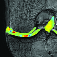
From the project abstract: The overall objective of the study is to determine the earliest stage of OA that can be detected using novel displacement-encoded MRI (deMRI) alone and in combination with conventional imaging and biomarker assays. In an effort to develop an imaging biomarker that noninvasively tracks structural degeneration in cartilage during OA progression, deMRI will be used for the measurement of mechanical strain in the interior of cartilage explants and intact joints in vivo. With the development of deMRI, the utility and translation of deMRI for diagnosing the early structural and mechanical changes to cartilage that may predispose the joint to deterioration but have not yet fully progressed to OA will be assessed.