Huang wins $1.8M NIH and $500k DARPA awards to advance super-resolution imaging
 On Aug. 1, 2016, Huang received the NIH Maximizing Investigators’ Research Award (MIRA) for New and Early-Stage Investigators. The five-year MIRA grant provides more than $1.8 million in funding, offering flexibility and stability to take on ambitious scientific projects which increases the likelihood of important breakthroughs. Huang’s research project is titled “Interferometric 3-D super-resolution imaging and structure and stoichiometry mapping in live cells.”
“Based on our recently developed ultra-high resolution 3D imaging system, we aim to make significant advancements in the directions of living cell and tissue imaging, which are two major roadblocks limiting the application range of modern super-resolution techniques,” Huang says. “This grant will enable us to create a new image modality for building dynamic structural models of large protein complexes in live cells. We are very excited to apply our developments to study cytokinesis, the final stage of cell division.”
 On Sept. 15, 2016, Huang also was honored with the DARPA Young Faculty Award. The DARPA award provides approximately $500,000 in funding over two years with the potential for an additional year of up to $500,000 under a DARPA Director’s Fellowship. Huang’s Young Faculty Award grant will provide support for Huang’s group to develop a novel nanoscope that significantly improves imaging depth and volume while minimizing light-induced toxicity.
“These super-resolution systems, or ‘nanoscopes’, produce an incredible amount of single-molecule data. It is a challenge and an opportunity for us to explore this new realm and to create new observation tools with this unique resource,” Huang says.
 “The NIH and DARPA awards facilitate two different research directions in my lab that are also synergistic parts,” he says. “I’m hoping we can combine elements of both projects to further push the envelope of super-resolution imaging and to shed light on biological processes that are otherwise hidden under the diffraction limit.”
The Rise of Super-Resolution Imaging
Super-resolution fluorescence microscopy gained prominence after its developers — Eric Betzig, Stefan Hell and William Moerner — received a 2014 Nobel Prize in chemistry. It has become a vital tool in cell biology research.
Recently, Huang and researchers from five other institutions developed an ultra-high resolution fluorescence nanoscope called “W-4PiSMSN,” capable of producing 3-D volumetric images at 10- to 20-nanometer resolution of an entire mammalian cell.
This new technology is detailed in an Aug. 11 research paper in the journal Cell.
Purdue’s Imaging Reputation
 Huang, who earned his PhD in physics from the University of New Mexico, came to Purdue in 2015 after his postdoctoral training in the Department of Cell Biology at Yale University. His transition to Purdue was partly inspired by the Provost’s Integrated Imaging Initiative cluster hire program.
The Integrated Imaging cluster was originally proposed by an interdisciplinary group of faculty from the colleges of science and engineering and focuses on the synergy between science and engineering to advance imaging research.
“The Integrated Imaging Initiative aims to exploit the synergy between science and engineering, algorithms and devices, sensors and applications,” Charles Bouman, Showalter Professor Electrical and Computer Engineering and Biomedical Engineering and chair of the recruiting committee, says. “It builds on strengths and leverages the imaging capabilities of Purdue’s engineering and science colleges and Purdue’s Discovery Park infrastructure.”
Huang looks forward to the future of his work in super-resolution imaging and believes the NIH and DARPA awards will significantly impact this emerging field of study.
“These awards provide me and my team with the necessary support to pursue our scientific goals, enabling me to spend more time doing the research I love,” Huang says.
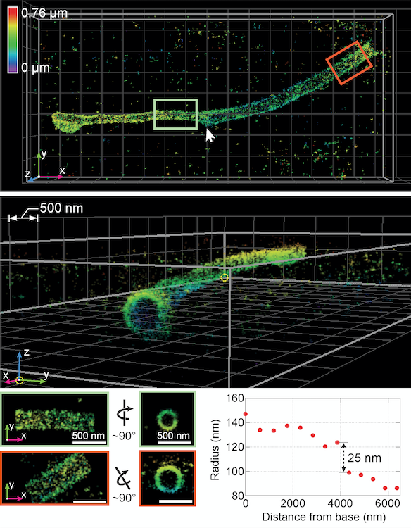
Image 1: Using a new 3-D super-resolution instrument, researchers captured this visualization of a "primary cilium," the antenna of the cell. (Image courtesy of Huang et al./Cell)
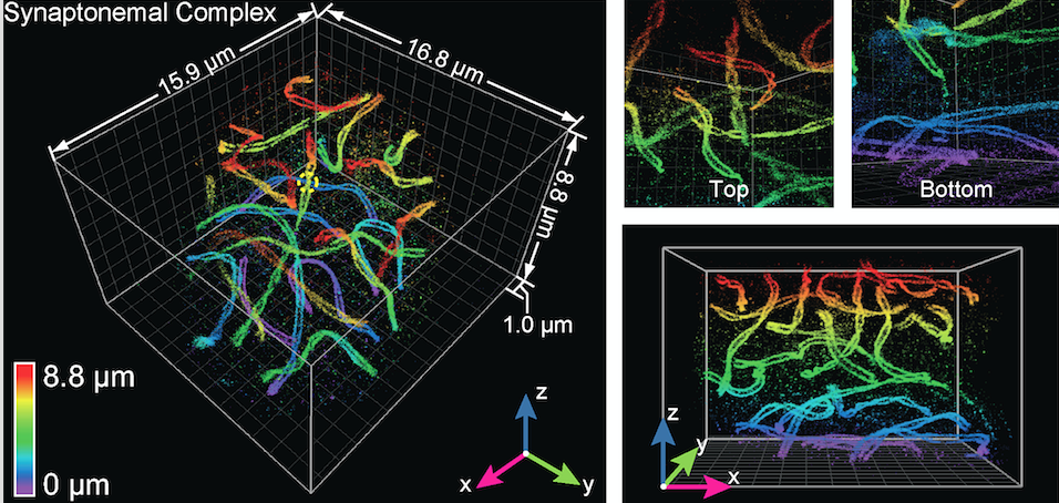
Image 2: Here, the technology was used to visualize a mouse spermatocyte, revealing with unprecedented clarity the "twisting paired lateral elements" of synaptonemal complexes, which link chromosomes together. (Image courtesy of Huang et al./Cell)