Ji-Xin Cheng's Non-Linear Microscopy Research Spotlighted
Cheng is collaborating with Stephen D. Miller, an immunologist at Northwestern University Medical School to study myelin sheaths that encase axons in the brain and spinal cord. CARS is an excellent tool to visualize axons with high resolution, allowing the researchers to observe the progression of diseases such as multiple sclerosis.
Another research project, in collaboration with Michael S. Sturek, a physiologist at the Indiana University School of Medicine, is combining CARS imaging with simultaneous sum-frequency-generation imaging to study lesions in atherosclerosis. By identifying specific lesions as containing unstable plaque, it is hoped that a majority of heart attacks could be prevented.
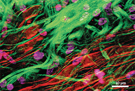
CARS and SRS techniques both utilize two pulsed laser beams that are tuned to match the frequency of a specific chemical or compound. The beams are aimed at a sample, and the resultant scattered light gives researchers images whose analysis benefits a broad range of applications, from medical diagnostics to pharmacokinetics to conversion of biomass to biofuels. These techniques can reduce imaging time from hours to seconds, with much higher resolution.
Additional information on Professor Cheng's research can be found at https://engineering.purdue.edu/BME/Research/Labs/Cheng