Catalysis and Biocatalysis
AI/ML Biocatalytic Pathway Design and Synthesis
Bioseparations and Biomolecular Characterization
Fermentation and Cell Culture
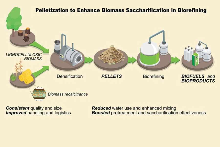
Lignocellulose Pellets: Resilient Feedstocks for Biomanufacturing