Medical Imaging Applications
Successfully registered medical images can provide medical workers with a quantitative and visual tool for studying diseases or injuries, and for helping with the detection and treatment in patients. They have also become more widely used in surgical planning. Medical image registration can be performed on images collected with different modalities and/or at different times. As a result, medical image registration has faced many challenges such as the large variations in resolution, differences in geometric and radiometric characteristics between modalities, and dynamic changes in anatomical structures due to disease progression for longitudinal studies. Parallel to medical image registration, challenges faced by the integration of multi-source and multi-temporal remotely sensed data acquired by earth observing satellites mandate the development of accurate, robust and automated registration procedures. Due to the similarities between these two applications, it is of great value to investigate the employment of satellite image registration techniques on medical imagery, which is the incentive behind this research.
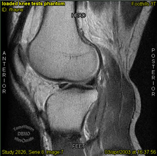
The research will concentrate on magnetic resonance (MR) image registration of knee joints that are captured at different physiologic loading conditions and at different times. Figure 1 shows an MR image of a loaded knee. The main objective is to utilize existing image registration methods for the development of new approaches that will enable accurate registration of MR images of both healthy and degenerated joints. More specifically, the Modified Iterated Hough Transform (MIHT) for Robust Parameter Estimation will be targeted with the use of higher order primitives (please refer to the research areas on Image Registration and Linear Features in Photogrammetry). The image registration accuracy, reliability and repeatability will be evaluated. This research is still in its preliminary stage but the expected results should allow a precise 3D reconstruction of the joints and can provide a better understanding of joint mechanics. Furthermore, this research can provide a validation on the reliability and accuracy of photogrammetric techniques.

Figure 1: Magnetic Resonance Image of a Loaded Knee Joint
