Materials Modeling
One of the primary goals of the work in the ACME2 lab is to introduce new lifing strategies into structural design practices. In today’s age of digital engineering, there are software tools to optimize engineering decisions for parts, yet the materials considerations are still based on trial-and-error approaches or the proverbial make-it and break-it method. In modern aircrafts with high demands for efficiency, materials are being pushed to their limit as defined by classical lifing approaches albeit without sacrificing safety standards. Our work introduces lifing strategies that are based on the material’s microstructure, while classical component life analysis is based on an overly conservative assessment of the material and assumes the material is constant across a part and any variation in properties is inherent to the material. By taking into account gradients in microstructure across a part due to the manufacturing process, the properties and associated life of the material can be determined at each location within the component. These quantifiable linkages between materials and properties have provided design and structures engineers with enhanced lifing analysis tools and the ability to reliably extend lifetimes of components. Moreover, these approaches allow the designer to tailor the microstructure at distinct sites within a component to achieve location specific properties.
Research themes:
- Microstructure based lifing approaches
- Location specific lifing
- Tailored materials properties
- Life extension analysis
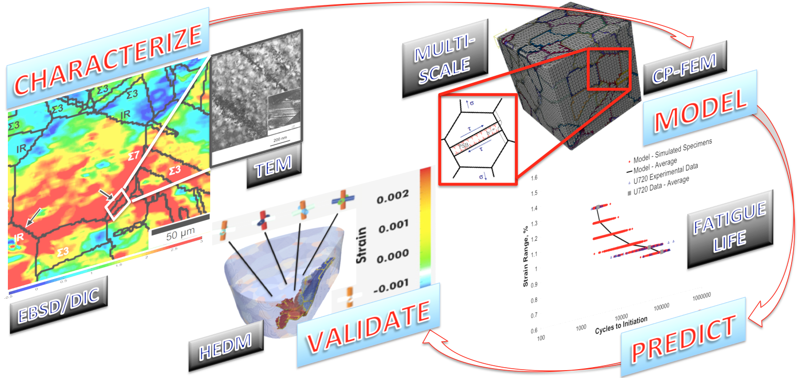
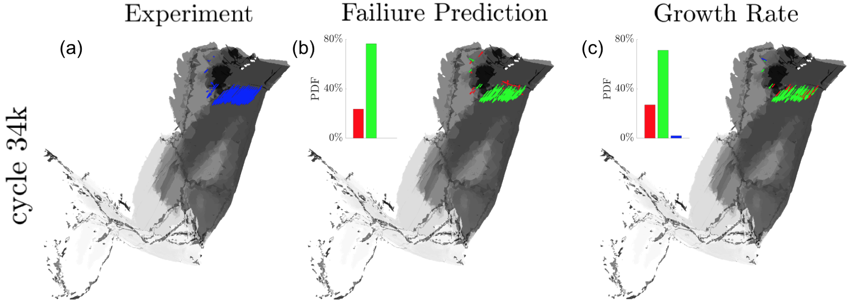
Specific modeling projects span a variety of failure mechanisms, such as fatigue crack initiation (via persistent slip bands, non-metallic inclusions, pores, etc.), fatigue crack propagation, fracture, creep, delamination, and interactions of these mechanisms. Moreover, our group has experience in modeling failure across a variety of structural engineering alloys, including polycrystalline metals (conventionally processed Ni-based superalloys, Al alloys, Ti alloys, and steels, but also in additive manufactured materials), ceramic matrix composites, and fiber-reinforced composites.