Seeing Inside Explosions With High Speed X-Rays
“Energetic materials are simply materials that have energy stored inside them, which can be released under certain conditions,” said Wayne Chen, Reilly Professor of Aeronautics and Astronautics & Materials Engineering. “This could include explosives used in mining operations, or the fuel that sends a rocket up into space. But the critical condition of when and how this energy gets released is still not completely understood.” As the principal investigator of a 5-year multidisciplinary project sponsored by the Air Force Office of Scientific Research, Chen and his team are conducting multiple tests to determine exactly what causes certain explosives to react, and then observing the reaction itself in unprecedented ways.
“Imagine you are transporting explosives to a mining site,” explained Chen. “Those materials have to be handled by people; they have to be transported by trucks. So there is vibration, and there are temperature shifts. We want these materials to release their energy when we want them to, and not accidentally! The more we can quantify about what causes energetic materials to react, the safer the process will be.”
So how do you test potent explosives without blowing up your lab? Chen’s solution is to start small, at what is called the mesoscale. Often they work with a single grain of energetic material, roughly the size of a sugar crystal. They suspend the crystal in a plastic binder to prevent it coming in contact with other crystals (creating a polymer-bonded explosive, or PBX). “It’s small enough that we can safely study it on campus,” said Chen, “but large enough that we can observe the reaction and extrapolate worthwhile results.”
 Chen’s team then subjects the crystals to various conditions, including heat, vibration, and low-speed impact. They know that it only takes a small “hotspot” to trigger a reaction, so what has the potential to cause these hotspots? “My area of expertise is impact damage of materials,” said Chen. “So we expose the samples to impact loading -- as if they had been dropped by someone handling them -- and observe exactly where the cracks form in the energetic materials.”
This creates two more big challenges. How do you observe a reaction that occurs in mere nanoseconds; and how do you look inside the particle to observe how the damage occurs? To solve these problems, Chen’s team utilized a unique technology: high speed X-ray cameras.
“We’ve all seen high-speed cameras that can shoot thousands of frames per second,” said Chen. “But to look through a sample at high speed, we need to use X-rays. And these X-rays need to be about a billion times more powerful than what you have at your dentist’s office! There are only three places in the world that can do this, and one of them is Argonne National Laboratory in Illinois.”
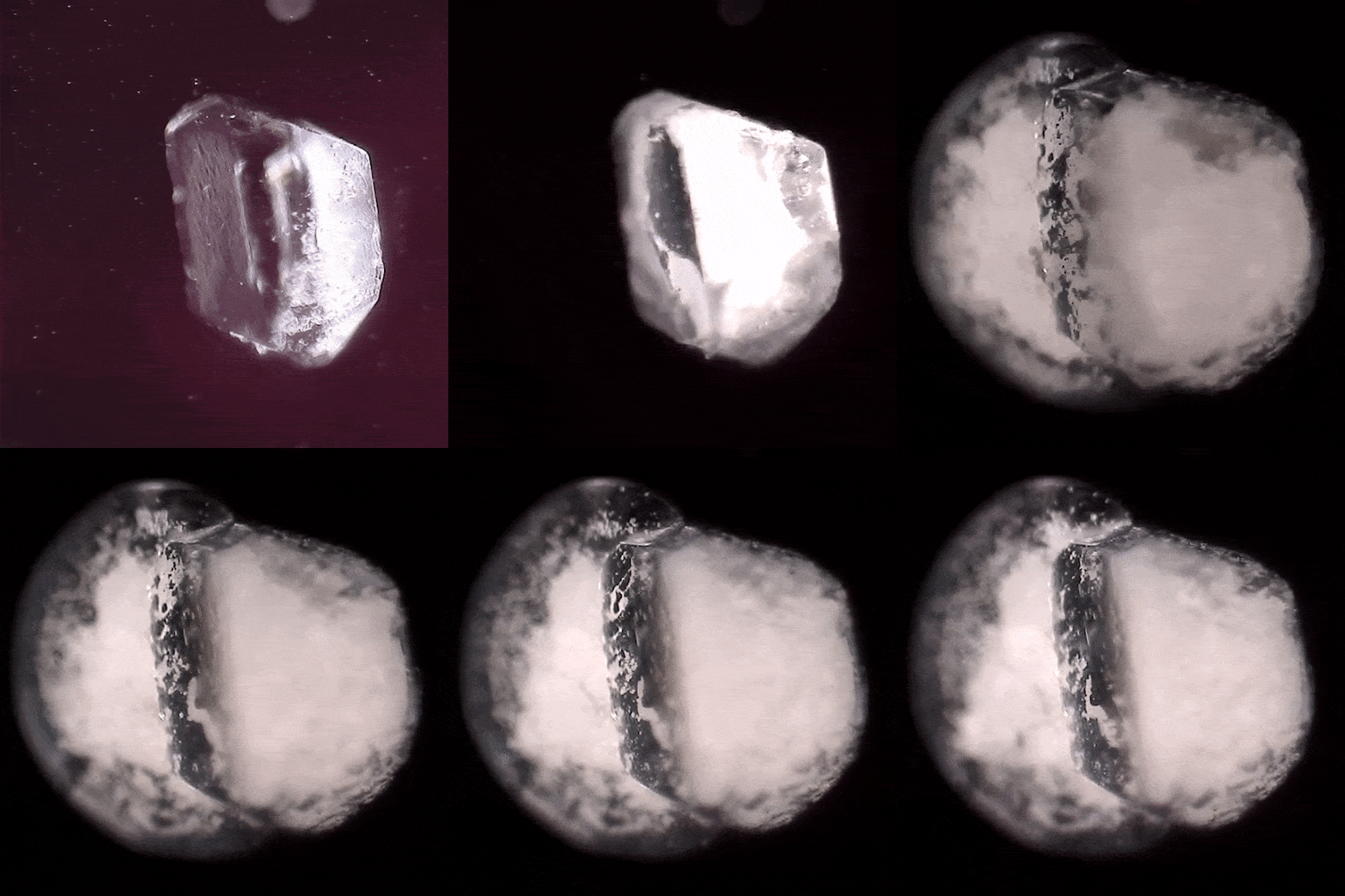
By using Argonne’s Advanced Photon Source synchrotron, the team have recorded groundbreaking high speed X-ray videos at five million frames per second, showing exactly what happens to an energetic crystal as it explodes. Chen described what they observed when exposing a sample to ultrasonic vibration: “The crystal changed its shape first, so we knew something was about to happen. Then it changed its phase, and became a pool of molten crystal, sloshing around. Then, it quickly became a gas, which is what happens when something explodes. But we were able to experimentally observe this process for the very first time.”
“That’s the best joy as a scientist,” said Chen. “We have seen something totally new. People have hypothesized about this for years, but no one has actually seen it until now! We’ve chased this for years, and finally we’ve recorded it.”
The team includes Steve Son, Alfred J. McAllister Professor of Mechanical Engineering, who creates the energetic material samples; Jeff Rhoads, professor of mechanical engineering, whose team focuses on vibration; Terry Meyer, professor of mechanical engineering, who measures the temperature response of the samples; Marisol Koslowski, professor of mechanical engineering, who uses computer models to measure the forces exerted on the particles; and Marcial Gonzalez, assistant professor of mechanical engineering, who uses particulate science to extrapolate the results to a larger scale.
“Purdue University is uniquely suited to do energetic materials research,” said Chen. “We have Zucrow Labs, the largest academic propulsion lab in the world, which is specifically set up to test rocket propellant and explosives. We have Herrick Labs, which is second-to-none in the country when it comes to vibration. And we have this team of talented faculty and graduate students with these unique skillsets. We’re all very excited, and also very privileged, because this is a critical phenomenon. We are serving our nation by making these materials safer.”
Writer: Jared Pike, jaredpike@purdue.edu, 765-496-0374
Source: Wayne Chen, wchen@purdue.edu, 765-494-1788
High speed X-ray phase contrast imaging of energetic composites under dynamic compression
Niranjan D. Parab, Zane A. Roberts, Michael H. Harr, Jesus O. Mares, Alex D. Casey, I. Emre Gunduz, Matthew Hudspeth, Benjamin Claus, Tao Sun, Kamel Fezzaa, Steven F. Son, and Weinong W. Chen
Fracture of crystals and frictional heating are associated with the formation of “hot spots” (localized heating) in energetic composites such as polymer bonded explosives (PBXs). Traditional high speed optical imaging methods cannot be used to study the dynamic sub-surface deformation and the fracture behavior of such materials due to their opaque nature. In this study, high speed synchrotron X-ray experiments are conducted to visualize the in situ deformation and the fracture mechanisms in PBXs composed of octahydro-1,3,5,7-tetranitro-1,3,5,7-tetrazocine (HMX) crystals and hydroxyl-terminated polybutadiene binder doped with iron (III) oxide. A modified Kolsky bar apparatus was used to apply controlled dynamic compression on the PBX specimens, and a high speed synchrotron X-ray phase contrast imaging (PCI) setup was used to record the in situ deformation and failure in the specimens. The experiments show that synchrotron X-ray PCI provides a sufficient contrast between the HMX crystals and the doped binder, even at ultrafast recording rates. Under dynamic compression, most of the cracking in the crystals was observed to be due to the tensile stress generated by the diametral compression applied from the contacts between the crystals. Tensile stress driven cracking was also observed for some of the crystals due to the transverse deformation of the binder and superior bonding between the crystal and the binder. The obtained results are vital to develop improved understanding and to validate the macroscopic and mesoscopic numerical models for energetic composites so that eventually hot spot formation can be predicted.
This research is funded by the Air Force Office of Scientific Research (AFOSR) under program managers Dr. Jennifer Jordan and Dr. Martin Schmidt through grant no. FA9550-15-1-0102.
