Purdue engineer, IU cardiologist collaborate to offer innovative tool and fresh hope for babies with heart defects
WEST LAFAYETTE, Ind. – When he was a medical student, none of Mark Payne’s textbooks told him that a cardiologist should consult an engineer before deciding on a patient’s treatment.
But today, as a pediatric cardiologist with Riley Hospital for Children at Indiana University Health and professor at IU’s School of Medicine, Payne wouldn’t consider investigating how to make the best use of an echocardiogram to evaluate congenital heart defects without involving engineers from Purdue University.
Pavlos Vlachos, the St. Vincent Health Professor of Healthcare Engineering and director of the Regenstrief Center for Healthcare Engineering, leads a Purdue team whose recent innovations are on track to be a game-changer for cardiac surgeons and to rewrite cardiology textbooks. Working with Payne, who contributes clinical and medical expertise, the two researchers are confident that they can help save babies’ lives by making echocardiograms — ultrasound image recordings of the heart — much more useful and surgery decisions far better informed.
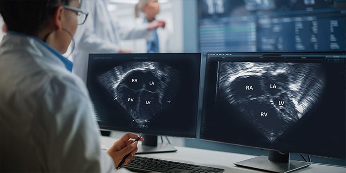
Vlachos’ tool, which is composed of four distinct elements, processes echocardiograms of fetal hearts in-utero using physics to analyze the function of blood flow within the heart and the heart tissue, and quantify its biomechanical performance. This detailed analysis, which has never been available before, provides information critical to planning highly complex heart surgeries and improving long-term outcomes for babies born with congenital heart defects — the single most prevalent birth defects in infants. The tool also is being studied at Children’s National Hospital in Washington, D.C..
“What makes our tool unique is that it combines medical imaging with advanced image-processing capability, physics and biomechanics,” Vlachos says. “It removes user input and guessing, and automates the process. It’s all objective and repeatable; it quantifies the function of the heart on the physics level with fundamental parameters — unlike what is currently used clinically, which often relies on heuristics, visual observations and manual measurements.”
Among the congenital heart problems that Vlachos, Payne and their teams are addressing is hypoplastic left heart syndrome (HLHS), a condition in which the left side of the heart is underdeveloped, necessitating surgery within 48 hours of birth. HLHS is responsible for 25% to 40% of all neonatal cardiac deaths and is always fatal without a series of complex corrective surgeries that begin urgently after birth. Treatments involving medicines and more surgeries continue throughout each patient’s life as the heart grows and ages.
“Right now, you can do an ultrasound and see the baby’s heart beating, but nobody can measure what we can about the function of that heart — or with the accuracy that we can,” Vlachos says. “There’s no other place, no other engineering capability or image-processing capability that can provide this kind of quantitative information.”
Payne’s enthusiasm about the technology matches Vlachos’. “We have lots of echocardiography studies over 30 years, and no one has pushed the envelope to say, ‘Well, what else can we get out of them?’ Pavlos is able to deconstruct the image and tell me how much energy is being lost as the blood flow goes through the heart, even in the fetal heart.”
Vlachos and Payne have just completed a report showing their teams’ recent work to test the tool. The research was funded by the Indiana Clinical and Translational Sciences Institute and a grant from the National Institutes of Health.*
Vlachos and his colleagues, Brett Meyers and Melissa Brindise, both former mechanical engineering doctoral research associates at Purdue, have intellectual property filings for the tool’s analysis algorithms.
Vlachos’ team and Payne, in collaboration with Children’s National Hospital, also are studying Tetralogy of Fallot, sometimes called blue baby syndrome, which is characterized by four distinct heart and blood vessel malformations that interfere with proper heart operation.
About the Regenstrief Center for Healthcare Engineering
Located in Discovery Park District at Purdue on the West Lafayette campus, the Regenstrief Center for Healthcare Engineering (RCHE) conducts research to improve the quality, accessibility and affordability of health care delivery through collaboration, partnerships and engagement. RCHE’s team is composed of researchers, staff and outreach advisors who collaborate with the health care community to develop science-based approaches to personalized care, match health resources with community needs and improve access to care among rural communities in Indiana and around the world.
*Study funding was provided by Grant Number UL1TR002529 from the National Institutes of Health, National Center for Advancing Translational Sciences, Clinical and Translational Sciences
Writer/Media contact: Amy Raley, araley@purdue.edu
Sources: Pavlos Vlachos, pvlachos@purdue.edu
Mark Payne, rpayne@iu.edu