Long-Term in-Vivo Monitoring of Neuromuscular Performance in Mice
The possibility of improving the quality of life of people living with neuromuscular disease motivates this study to develop a closed-loop electrical stimulation therapy delivery system.
Neuromuscular diseases are broadly characterized by impaired function of muscles, peripheral nerves, or the neuromuscular junction. These diseases manifest themselves with a range of symptoms including muscle weakness, atrophy, and aberrant activity such as fasciculations and fibrillations. Electrophysiology is one tool we can use to study neuromuscular disease mechanisms in animal models. Longitudinal electrophysiology or behavioral studies of these models typically involve the implantation of a transcutaneous adapter and subsequent tethering of a mouse, which is the premier mammalian model for most human diseases. This strategy resolves the limitations of performing electrophysiology in an anaesthetized mouse?namely, the suppression of spontaneous, motor evoked activity?while permitting the mouse to behave somewhat freely. However, the tethered system places restrictions on the testing environment and the types of activities the mouse can perform. Additionally, tethered systems are not conducive to continuous, 24-hour monitoring, which is desirable when tracking spontaneous or unpredictable bioelectric activity.
The fully implantable solution being investigated for longitudinal electrophysiology in mice?the Bionode?is a wireless system that eliminates the need for tethering during behavioral or bioelectric monitoring. The device is implanted subcutaneously and allows for continuous monitoring of spontaneous activity. The Bionode is powered wirelessly using electromagnetic fields within a closed environment, which eliminates the need for an on-board battery. This system allows us to continuously observe activity within the muscles and peripheral nerves, which can be used to study neuromuscular disease mechanisms or evaluate the effectiveness of new treatments and therapies for these diseases.
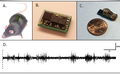
In the figure above, part A provides a representation of the Bionode device placed subcutaneously in the mouse with electrodes recording from a muscle target in the hindlimb. Part B of the figure shows the latest iteration of the Bionode system designed to collect and wirelessly transmit bioelectric information. Part C of the figure displays the Bionode device with power coupling coils, which would be entirely sealed in medical grade epoxy prior to implantation. Part D shows electromyography (EMG) collected from a mouse for familial muscular dystrophy showing aberrant muscle fasciculations.
- Project Researchers: Rebecca Bercich