'Fingerprinting' cell metabolism points toward study of obesity, diabetes
September 8, 2014
|
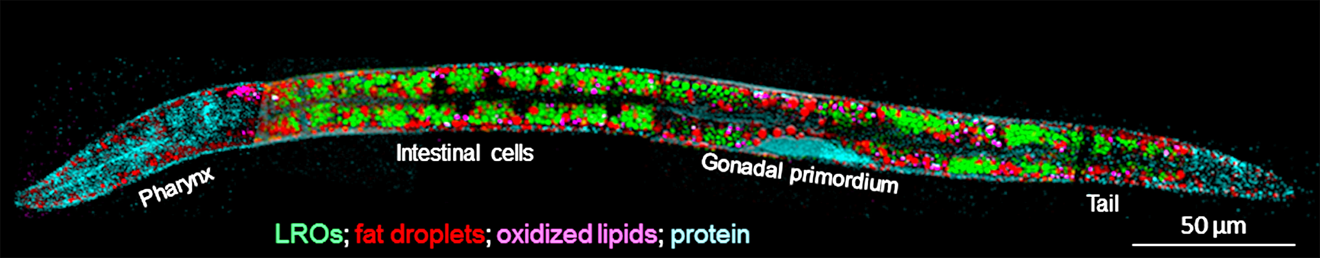
Researchers have used a new imaging platform to map lipid metabolism in the living cells of a transparent round worm called C. elegans. Findings point toward further studies into the impact of diet and the role of insulin in obesity, diabetes and longevity in animals and humans. (Ji-Xin Cheng/Purdue University image) |
WEST LAFAYETTE, Ind. – Researchers have shown how to use a new imaging platform to map lipid metabolism in living cells, discovering specifically where cholesterol is stored and pointing toward further studies in obesity, diabetes and longevity.
The imaging approach makes it possible to not only quantify the storage of cholesterol - a type of lipid - but also the "desaturation" and oxidation of lipids, which may reduce the ability of cells to use insulin, said Ji-Xin Cheng, a professor in Purdue University's Weldon School of Biomedical Engineering and Department of Chemistry.
The research was conducted with live roundworms called C. elegans, which are used extensively to study the impact of lipid metabolism on aging and disease.
"This animal is transparent, so we can see what's happening in real time," Cheng said. "We can follow the metabolism inside the animal."
Findings are detailed in a research paper appearing this month in the journal Angewandte Chemie. The paper was authored by Purdue postdoctoral researchers Ping Wang, Bin Liu and Delong Zhang; University of Massachusetts (UM) Medical School doctoral student Micah Y. Belew; Heidi A. Tissenbaum, a professor in the UM Medical School's Program in Gene Function and Expression/Program in Molecular Medicine; and Cheng.
The researchers used an imaging platform called hyperspectral stimulated Raman scattering microscopy, which can identify and track certain molecules by measuring their vibrational spectrum with a laser, a sort of spectral fingerprint.
Whereas conventional Raman microscopes may take hours to get results, the new method works at high speed, enabling researchers to measure changes in real time in live animals.
"The advantage of being able to observe what is happening in real time in a live tissue is that you can follow the same cell over time, just like following the same person over time to track a patient's health," Cheng said.
Being able to map the degree of lipid unsaturation and oxidation and cholesterol storage inside living cells will make possible further studies into the impact of diet and the role of insulin in obesity, diabetes and longevity in animals and humans.
"This work is a result of a successful collaboration between our labs. The new technology has allowed us to observe changes in lipids in a live animal," Tissenbaum said. "Most analyses of lipids examine the composition of whole animal extracts. Now, we can observe changes in the different tissues in real time. Now, we can answer the important questions of how lipid stores change in response to diet and age."
Ordinarily, cells have to be processed before they can be analyzed, which rules out the possibility of studying living cells.
"The promise of doing in vivo imaging is to learn where the key chemicals and molecules are, what their dynamics are and how they communicate to each other," Cheng said.
Findings revealed that cholesterol is stored in lysosome-related organelles (LROs), compartments inside cells containing digestive enzymes.
"The cell is like the human body because inside there are many compartments, which are like organs," Cheng said. "Our results reveal for the first time that lysosome related organelles in intestinal cells are sites for storage of cholesterol in C. elegans."
The work is supported by the National Institutes of Health and the William Randolph Hearst Foundation.
The research is ongoing.
"One thing we will do next is to overfeed C. elegans with glucose and then study the role of diet in lipid metabolism and cholesterol storage," Cheng said.
Writer: Emil Venere, 765-494-3470, venere@purdue.edu
Source: Ji-Xin Cheng, 765-494-4335, jcheng@purdue.edu
Related website:
Department of Basic Medical Sciences: http://www.vet.purdue.edu/bms/
Note to Journalists: An electronic copy of the research paper is available from Emil Venere, 765-494-4709, venere@purdue.edu
ABSTRACT
Imaging Lipid Metabolism in Live Caenorhabditis elegans Using Fingerprint Vibrations**
Ping Wang, Bin Liu, Delong Zhang, Micah Y. Belew, Heidi A. Tissenbaum *, Ji-Xin Cheng*
Purdue University
Quantitation of lipid storage, desaturation, and oxidation in live C. elegans has been a longstanding obstacle. By hyperspectral stimulated Raman scattering imaging and multivariate analysis in fingerprint vibration region, we present a platform that allows quantitative mapping of fat distribution, degree of fat unsaturation, lipid oxidation, and cholesterol storage in vivo in whole worm. Our results reveal for the first time that lysosome related organelles in intestinal cells are sites for storage of cholesterol in C. elegans.
