New imaging tech promising for diagnosing cardiovascular disease, diabetes
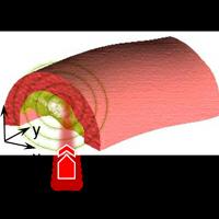
Professor Ji-Xin Cheng and his team members developed a new imaging technology to diagnose cardiovascular disease and other disorders by measuring ultrasound signals from molecules exposed to a fast-pulsing laser.
The new method could be used to take precise three-dimensional images of plaques lining arteries, said Cheng.
Other imaging methods that provide molecular information are unable to penetrate tissue deep enough to reveal the three-dimensional structure of the plaques, but being able to do so would make better diagnoses possible, he said.
"You would have to cut a cross section of an artery to really see the three-dimensional structure of the plaque," Cheng said. "Obviously, that can't be used for living patients."
The imaging reveals the presence of carbon-hydrogen bonds making up lipid molecules in arterial plaques that cause heart disease. The method also might be used to detect fat molecules in muscles to diagnose diabetes and for other lipid-related disorders, including neurological conditions and brain trauma. The technique also reveals nitrogen-hydrogen bonds making up proteins, meaning the imaging tool also might be useful for diagnosing other diseases and to study collagen's role in scar formation.
"Being able to key on specific chemical bonds is expected to open a completely new direction for the field," Cheng said
Findings are detailed in a paper to be published in Physical Review Letters and expected to appear in the June 17 issue. The findings represent the culmination of four years of research led by Cheng and doctoral student Han-Wei Wang.

